The pancreas is a long organ, measuring 12-15 cm, located against the back wall of the abdominal cavity, corresponding to the second lumbar vertebra, and situated within the duodenum. Therefore, when the pancreas is diseased, it can be easily misdiagnosed as a duodenal condition.
There are two types of pancreatitis:
Acute Non-Necrotizing Pancreatitis
 |
|
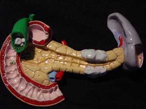
Pancreas Model (Image: pancreas) |
Acute pancreatitis is often seen in patients with biliary stones, such as gallstones, intrahepatic bile duct stones, and common bile duct stones (choledocholithiasis), as well as in cases of ascariasis. Therefore, patients presenting with renal colic suspected to be due to biliary stones or ascariasis must be carefully evaluated for acute pancreatitis.
The primary symptom of acute non-necrotizing pancreatitis is sudden, severe pain in the right upper quadrant that radiates to the back and shoulder. The pain can sometimes feel like “a kick in the back” as in bile duct obstruction with worms, accompanied by fever, chills, and tenderness when palpating the costovertebral angle, especially on the left side. The pain typically lasts for a few hours before gradually subsiding. Patients may feel fatigued, and sometimes another episode of pain may occur.
Laboratory tests typically show elevated white blood cells, amylase levels, bilirubin, and blood urea. Ultrasound is valuable for diagnosis but may sometimes be challenging due to gas and fluid accumulation in the intestine obscuring the image of the pancreas.
Acute Hemorrhagic Pancreatitis
Unlike acute non-necrotizing pancreatitis, acute hemorrhagic pancreatitis is less common among patients with gallstones but often occurs due to bile duct obstruction from worms and infections of the biliary tract caused by microorganisms.
Patients often present with noticeable shock. The main symptom is pain in the upper abdomen, radiating from the left costal margin to the back. The pain is continuous with moderate intensity, often interrupted by severe episodes, accompanied by nausea and vomiting. Vomiting may include food, yellowish fluid, diarrhea, and blood-stained stool. Patients may appear panicked and sweaty. In the early hours of the condition, hypertension may occur, but blood pressure typically drops afterward. Abdominal examination reveals tenderness, especially in the upper abdomen and the left costovertebral angle. In these cases, misdiagnosis with gastric perforation is common.
An unprepared X-ray should be taken to check for air-fluid levels. The radiograph may show distended intestinal loops near the pancreas, such as the duodenal loop and the proximal jejunum. Bruising may be seen around the umbilical area. Occasionally, pleural effusion may be present. Ultrasound images can show edema of the pancreas, with surrounding fluid accumulation.
Amylase levels are typically elevated within the first 12-18 hours. If blood sugar levels and particularly amylase remain elevated for seven consecutive days, this indicates a poor prognosis, and an increase after two weeks suggests the possible formation of a pancreatic cyst or ascites due to pancreatic fluid leakage. Additionally, urine tests may reveal the presence of albumin, red blood cells, and white blood cells.
Treatment
In cases of acute pancreatitis, patients require intensive treatment, including fluid resuscitation; pain management (morphine should be avoided as it can cause spasms of the Oddi sphincter); shock management (if present); and antibiotic therapy to prevent infection, with surgical intervention if necessary.
Prevention of Acute Pancreatitis
To prevent the disease, patients should undergo regular stool examinations for deworming as advised by their physician; avoid excessive alcohol consumption. Necrotizing pancreatitis is a very dangerous condition, and vigilance is necessary for early diagnosis and timely management to reduce mortality. When biliary stones and gallstones are detected, definitive treatment is required.


