Research Team at Peking University Achieves Breakthrough in Multiphoton Microscopy with Successful Imaging of Deep Brain in Mice.
The microscope, weighing only 2.17g, has successfully captured stable images of the cortex and hippocampal neurons in freely moving mice, a promising method that could unlock the mysteries of the human brain in the future, according to a study published in the journal Nature Methods on February 24th.
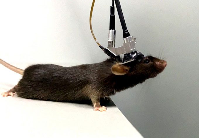
A mouse wearing the three-photon microscope developed by Peking University exploring its surroundings. (Photo: Xinhua)
The research team, led by Cheng Heping, Director of the National Biomedical Imaging Center at Peking University, has been developing this device for many years.
They created the first miniaturized two-photon microscope weighing 2.2g in 2017, capturing dynamic images of neuron and synapse activities in the cortex of freely moving mice.
After years of development, the team upgraded the two-photon microscope to expand the imaging field by 7.8 times, enabling it to capture three-dimensional images of functional signals from cortical neurons. The new three-photon microscope offers much greater imaging depth compared to previous miniaturized multiphoton microscopes.
This device can penetrate the entire cortex and the optic tract (a thick bundle of nerves beneath the cerebral cortex) of freely moving mice, recording images of calcium activities in the hippocampal region at depths of up to 1.2 mm, a significant challenge for neuroscientists worldwide.
Calcium activity serves as an indicator of neuronal activity and can be monitored when combined with fluorescent molecules. However, light scattering in brain tissue, especially in the optic tract beneath the cortex, hinders the penetration of fluorescence, limiting imaging depth.
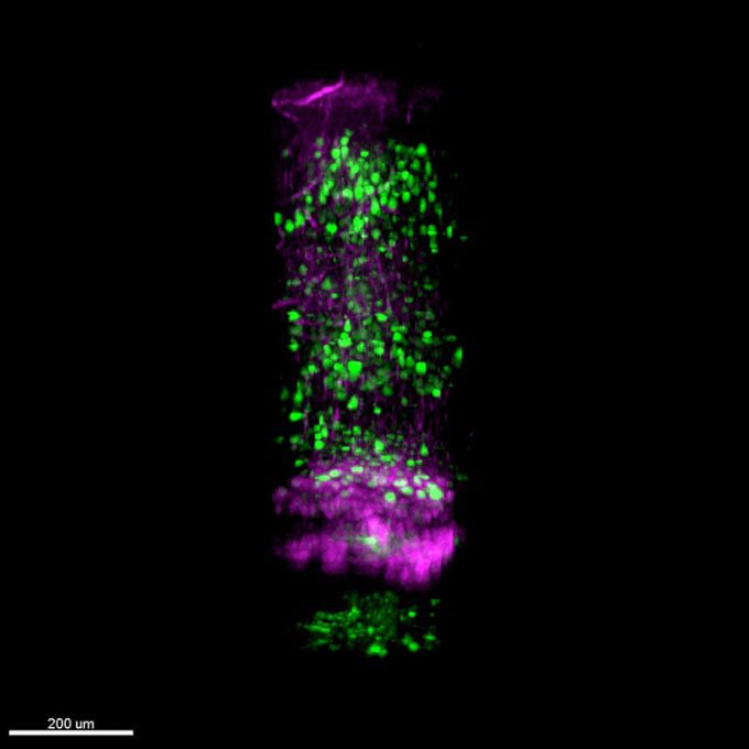
Images captured by the three-photon microscope show the cortex and hippocampal region in the deep brain of mice, with green spots indicating the fluorescent calcium signals of neurons. (Photo: Xinhua).
The hippocampus lies beneath the cortex and the optic tract. Previous miniaturized multiphoton microscopes worldwide have been unable to capture images of these areas non-invasively, according to Zhao Chunzhu, a member of the research team at Peking University.
The three-photon microscope developed by Cheng’s team successfully captured images in the deep brain of mice thanks to an innovative optical configuration that maximizes the collection efficiency of scattered fluorescence.
According to the research, the microscope also boasts the advantage of low optical toxicity, preventing fluorescence bleaching or early aging while observing neuronal activities over extended periods.


