Italian researchers are applying modern medical technology to uncover the mysteries surrounding an Egyptian mummy.
An ancient Egyptian mummy was transferred on June 21 from the Archaeological Museum in Bergamo to the Policlinico Hospital in Milan, where experts will use computed tomography (CT scan) technology to illuminate his life and burial customs from nearly 3,000 years ago.
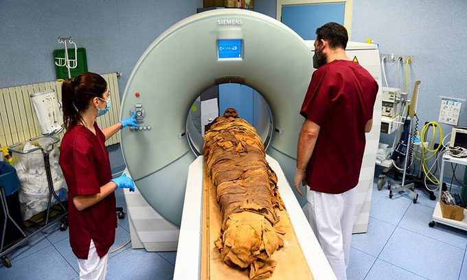
Medical staff scanning the nearly 3,000-year-old mummy. (Photo: Reuters).
“This is a type of virtual autopsy. We can create 3D images to study the overall mummy, allowing us to reconstruct this person’s life and death. We can also measure height and reassess gender based on pelvic analysis,” said Sabina Malgora, Director of the Mummy Project Research.
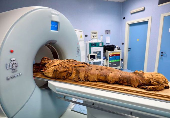
CT scan technology allows for 3D imaging to study the mummy comprehensively. (Photo: Reuters).
In addition to CT scanning, medical staff are also conducting chemical and physical analyses to understand what substances were used in the embalming process.
“Mummies are truly a biological museum. They are like time capsules. Studying ancient diseases and injuries is crucial for modern medical research,” Malgora added.
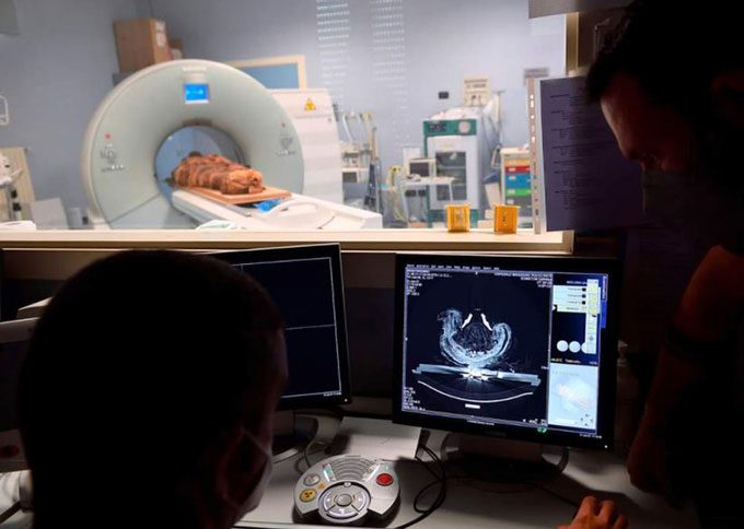
The research team viewing CT scan images of the mummy on a computer screen. (Photo: Reuters).
The inscriptions on the coffin (dating from around 900 BC to 800 BC) reveal that the mummy belonged to an ancient Egyptian priest named Ankhekhonsu, which means “The living God Khonsu.”
The results of the CT scan may confirm whether this is accurate. In April, a mummy believed to be the remains of a male priest was scanned by Polish scientists, who discovered that it was actually the body of a young woman in her 20s who was pregnant.


