Nasopharyngeal fibroma is a benign tumor that can spread widely, destroy bone tissue, significantly deteriorate health, and pose a life-threatening risk to patients if not detected and treated promptly.
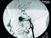 |
| Image of nasopharyngeal fibroma (Photo: TTO) |
Historically, individuals with nasopharyngeal fibroma were described with horrifying features such as bulging eyes, a widened nose, and cheeks swollen like pebbles. Today, we no longer encounter such severe cases as the disease is detected earlier.
Nasopharyngeal fibroma is a common condition in adolescence, particularly among males aged 15-25 in rural areas. It is believed that the development of nasopharyngeal fibroma is linked to hormonal changes during puberty.
The fibroma adheres closely to the base of the skull in the nasopharyngeal region and infiltrates adjacent structures, forming attachments in the sphenoid region, around the posterior nasal aperture, and the medial pterygoid plate. The tumor is firm, lobulated, and has a stalk. Its size varies depending on the duration between the onset of the disease and the surgical removal. Microscopic examination of the tumor reveals numerous blood vessels and scattered pools of blood along with fibrous tissue, forming a network under the mucosa that envelops the entire tumor.
Progression of the Disease
The disease progresses silently with symptoms of increasing unilateral nasal obstruction, which later expands to bilateral nasal obstruction in advanced stages. Continuous nasal discharge gradually increases, resulting in both nostrils becoming wet and congested with mucus. Occasional nosebleeds may occur, initially manageable without intervention but later requiring medical assistance to control. Accompanying these symptoms are signs of ear fullness and hearing loss due to the tumor compressing the Eustachian tube area. Patients may appear pale and fatigued.
Examination of the nasal cavity reveals a pink tumor obstructing the posterior nasal aperture; as it grows larger, it fills the nasal cavity, pushing against and narrowing the opposite nasal cavity. Examination of the throat and posterior rhinoscopy shows the soft palate is swollen, pushed forward and downward, or overly large, extending into the throat. A gentle palpation assesses whether the tumor is hard or soft, large or small, and mobile or fixed.
If allowed to develop naturally, the tumor will fill the nasal cavity, maxillary sinuses, sphenoid sinuses, and eye sockets, breaking through the hard palate, resorbing the upper jawbone, and causing subcutaneous swelling. The tumor may invade the cranial cavity, damaging cranial nerves, leading to blindness, swallowing difficulties, and loss of smell. Patients with untreated nasopharyngeal fibroma may die from bleeding and intracranial complications that cause increased intracranial pressure.
A definitive diagnosis is made through clinical examination: a pink tumor at the posterior nasal aperture in male patients aged 15-25, with nasal bleeding, should be referred to an ENT specialist for CT scanning and angiography for accurate assessment.
Treatment of the Disease
Surgery is the primary treatment method; if detected early, the tumor can be removed through an endoscopic approach, which does not leave scars around the nose as seen with conventional surgery for larger tumors. In cases where the tumor is too large for surgical intervention, radiation therapy combined with hormonal treatment may be recommended to shrink the tumor.


