Individuals infected with Trichinella spiralis due to consuming undercooked animal meat can experience symptoms such as intestinal inflammation and hemorrhage. In severe cases, it may lead to dangerous complications like pneumonia and encephalitis.
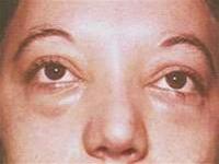 |
| Eye swelling symptom due to Trichinella infection (Image: TTO) |
Trichinosis is an infection caused by adult Trichinella worms and their larvae. The first identification of these worms was made by a first-year medical student named Jame Paget on February 6, 1835. To date, over a dozen species of Trichinella have been documented, with some common species including: Trichinella spiralis, which parasitizes livestock and can cause illness in humans; Trichinella britovi, which primarily infests wild boars and horses; and Trichinella nelsoni, which primarily infects carnivorous animals.
Trichinosis was discovered in 1835, and it was quickly found that Trichinella worms are present worldwide, with a higher prevalence in the Northern Hemisphere.
In the past 20 years, despite active prevention efforts, cases of trichinosis have been increasingly reported in many countries, and outbreaks have even occurred. Many scientists believe that trichinosis should be classified as a notifiable disease.
Ecological changes due to overexploitation are occurring globally, yet the lack of a stringent monitoring system has led to an increase in infection rates. According to a report from WHO, in March 2001, an outbreak of trichinosis occurred in Italy, affecting thousands of individuals, with 50% of pigs investigated during this outbreak testing positive for Trichinella.
Life Cycle of Trichinella
In the host, Trichinella can exist in both adult and larval forms. Adult worms reside in the intestines, while the larvae inhabit muscle tissues. Adult male worms measure only 1.4-1.6 mm, while females are longer, ranging from 3-4 mm. The hosts for Trichinella include humans and domesticated animals such as pigs, cats, and horses, as well as wild animals like foxes, bears, wild boars, wolves, and rats. Hence, trichinosis has both natural and human-associated reservoirs.
Human trichinosis is primarily associated with Trichinella outbreaks in nearby animal populations. Individuals become infected by consuming undercooked meat from animals that harbor Trichinella cysts. Once ingested, the larvae are released from the cysts in the stomach and migrate to the small intestine within 1-2 hours.
Here, after 24 hours, the larvae develop into adult worms and invade the intestinal mucosa. By days 4-5 and lasting from 10-30 days, the female worms release larvae into the lymphatic vessels. The larvae travel through the lymphatic system to the right heart, lungs, left heart, and then to skeletal muscles, diaphragm, and tongue, where they develop into cysts capable of causing infection. Trichinella cysts can survive in muscle tissue for about 20 years while still retaining their infectious potential.
Symptoms of Trichinella Infection
The incubation period for trichinosis ranges from 10 to 25 days; a shorter incubation period often correlates with a more severe illness. As the larvae invade the body and mature in the intestines, inflammation and hemorrhage occur, causing abdominal pain and diarrhea. The clinical manifestations of trichinosis encompass four primary symptoms:
– Eye swelling is a characteristic sign of the disease and may be accompanied by conjunctival edema and bleeding, sometimes leading to swelling of the entire head or even the neck and upper limbs.
– Muscle pain: Pain occurs during deep breathing, coughing, chewing, swallowing, or defecating, affecting both facial and neck muscles.
– Fever: Typically, body temperature gradually rises, peaking after 2-3 days. In mild cases, the illness may progress with a low-grade fever.
– Eosinophilia: Eosinophil levels rise during the initial days of infection, potentially detectable even during the incubation period, peaking in the third week of the illness.
In addition to the symptoms listed, various skin rashes resembling urticaria may appear. In severe cases, complications often arise in the third or fourth week, such as myositis, pneumonia, and encephalitis, which can be fatal. Depending on the severity of the infection, the mortality rate ranges from 6% to 30%.
Treatment and Prevention
Diagnosis of trichinosis is based on specific clinical symptoms and laboratory findings. Confirmatory diagnosis requires additional testing results. Blood tests typically show increased eosinophils; in the early stage of the disease, adult worms may be found in the stool, while larvae can be identified in biopsy specimens during the acute phase. Immunological responses such as complement fixation, agglutination, fluorescence immunoassays, and ELISA may also be utilized.
It is crucial to differentiate trichinosis from other conditions such as serum sickness, dermatitis, myositis, allergic bronchitis, pneumonia, and influenza.
Depending on the severity of the disease and the patient’s condition, medications like praziquantel, mebendazole, or albendazole may be prescribed by a physician.
Given that Trichinella has both natural and human-associated reservoirs, preventing this disease is quite challenging. In European and American countries, routine meat inspections have detected Trichinella infections in pork at rates ranging from 0.4% to 2.5%, with some areas reporting up to 10%. Therefore, the best preventive measure is to conduct thorough inspections for Trichinella in meat and to avoid consuming undercooked meats.


