Currently, there is a phenomenon where everyone fears being diagnosed with osteoporosis and rushes to measure bone density (BD). However, we need to recognize that any diagnostic method has its limitations and can lead to increased costs in terms of time, money, and additional anxiety and fear.
Why is it necessary to measure bone density for osteoporosis diagnosis?
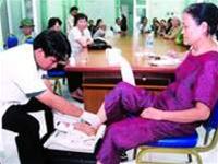 |
| Measuring bone density for patients at the Geriatric Institute (Photo: TTO) |
Osteoporosis (OP) is a pathological condition of the skeletal system characterized by a decrease in bone mass (i.e., a reduction in bone mineral density, primarily calcium), combined with microstructural deterioration of bone tissue, making bones fragile and increasing the risk of fractures. In theory, diagnosing OP requires the simultaneous evaluation of two factors: bone mineral density and bones’ microstructure.
There are various methods for diagnosing OP. The first involves direct assessments of bone structure (through bone biopsy) or indirect assessments (using nuclear magnetic resonance to evaluate bone structure in three-dimensional space). Bone biopsy for OP diagnosis is painful and can only be performed on the iliac bone, making it impractical for widespread use. Nuclear magnetic resonance is very expensive and has not yet been applied in clinical practice, remaining limited to scientific research.
Clinical diagnosis often occurs late because OP progresses silently, without symptoms, and only manifests in late stages when complications arise. These include spinal pain due to vertebral fractures, accompanied by height loss, kyphosis, or fractures of the femur, wrist, pelvis, or ribs. These complications require long-term, costly treatment, with a high mortality rate.
X-ray diagnosis also only allows for detection when bone density has lost 30-50%. Therefore, X-rays are not recommended for definitive OP diagnosis. Biochemical tests to assess bone turnover (the process of bone formation and resorption) are very costly and do not permit evaluation of bone density or prediction of fracture risk. For this reason, the use of bone turnover markers for diagnosis and treatment monitoring is not recommended.
Currently, the World Health Organization (WHO 1994) stipulates the use of bone mineral density measurement for diagnosing OP. Measuring bone density also allows for early diagnosis of OP, before severe complications like fractures occur. Thus, bone density measurement (BDM) is considered the gold standard for diagnosing OP.
Who should have their BDM measured?
Firstly, those at high risk of OP should have their BDM measured. This includes older adults, Caucasians or Asians, individuals with a history of fractures after age 40-45, a family history of fractures or OP, prolonged immobilization or low physical activity, or low body weight (BMI).
Some authors suggest that women who do not use hormone replacement therapy in the first five years after menopause are at a higher risk of OP compared to others. It is not necessary to measure BDM for all postmenopausal women. BDM should only be measured in women who experience early menopause before age 40 or those who have normal menopause but a history of conditions that may lead to secondary OP (prolonged hypogonadism, untreated progressive hyperthyroidism, adrenal hyperplasia, and primary hyperthyroidism).
Additionally, BDM should be measured in individuals showing clinical signs of suspected vertebral compression fractures, height loss, kyphosis, or fractures of the arms or legs occurring without significant trauma.
When should BDM be repeated?
If there is no treatment indication after measuring BDM, it should only be repeated after 2-3 years, or even up to 4 years. To detect a 2-3% change in BDM, it may take 1-1.5 years if the error is 1%, and 5 years if the machine error is 5%. When re-measuring, it is important to use the same machine at the same location.
In cases of treatment, BDM may be repeated at the lumbar spine since the effects of medication at this site are quite pronounced.
What other factors should be considered besides BDM?
Many other factors contribute to fractures besides just BDM. These include risk factors for fractures that can easily lead to femoral fractures such as poor vision, decreased mobility due to immobilization, unsuitable environments (any objects that cause falls at home), decreased muscle strength (difficulty climbing stairs, reduced knee extension strength, or quadriceps strength), use of sedative medications, or factors that affect balance. The type of fall, height, and length of the femur are also risk factors independent of BDM.
In summary, BDM should only be measured in individuals at high risk for OP or suspected of having it. Only then will the measurement of BDM be fully effective.


