The scientists who created this monkey embryo are the same individuals who successfully cloned the first two monkeys in 2018 using somatic cell nuclear transfer techniques.
Every biology textbook you have ever studied states that an embryo from sexually reproducing organisms is the result of the fertilization process between a female’s egg and a male’s sperm. This means that without either of the two, no embryo can be produced.
However, this premise has now been permanently altered. In a recent study published in the journal Cell Stem Cell, Chinese scientists have for the first time created monkey-like structures resembling embryos without the need for either eggs or sperm.
These embryos were instead cultivated directly from the stem cells of crab-eating macaques (Macaca fascicularis).
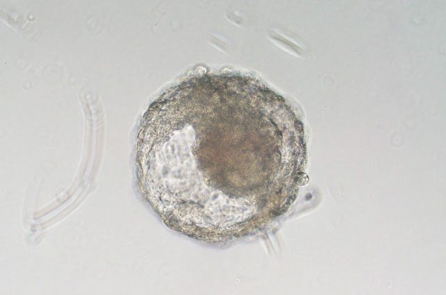
This is an artificial embryo of the Macaca fascicularis monkey developed by Chinese scientists.
“This is a significant milestone in the field of stem cell-derived embryo modeling,” stated Alejandro De Los Angeles, a biologist from the University of Oxford.
The creation of these synthetic monkey embryos will aid scientists in unraveling many mysteries related to the embryo formation and pregnancy processes in humans, without facing the ethical constraints associated with using real human embryos.
This research is expected to advance several fields, including infertility treatment, miscarriage prevention, and screening for congenital diseases during embryo development.
How were these monkey embryos created without eggs and sperm?
The new study was conducted by Dr. Zhen Liu, a developmental biologist at the Institute of Neuroscience, Chinese Academy of Sciences, Shanghai (CAS), and Dr. Qiang Sun, a neuroscientist and director of the Non-Human Primate Research Facility at CAS.
If you recall, this duo was responsible for the successful cloning of two Macaca fascicularis monkeys named Zhong Zhong and Hua Hua in 2018. These were the first monkeys born using somatic cell nuclear transfer, the same method used to clone Dolly the sheep, the first cloned mammal in 1996.
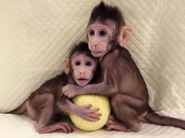
Zhong Zhong and Hua Hua, the cloned crab-eating monkeys created by the same research team in 2018.
In the 2023 study, Dr. Liu and Dr. Sun continued to explore the crab-eating macaque. This species, native to Southeast Asia, is often seen on beaches as they attempt to catch crabs, hence the name crab-eating monkeys.
Crab-eating macaques share habitats with humans and, due to their close genetic relationship with us, are frequently used in biomedical experiments and research.
For this study, Dr. Liu’s team collected embryonic stem cells from crab-eating macaques and exposed them to a mixture known as growth factors. Growth factors consist of nutrients and various molecules capable of stimulating stem cells to develop into different cell types.
Theoretically, stem cells can develop into any cell in the body. With the right formula, the Chinese scientists were able to “persuade” these cells to transform into embryonic cells.
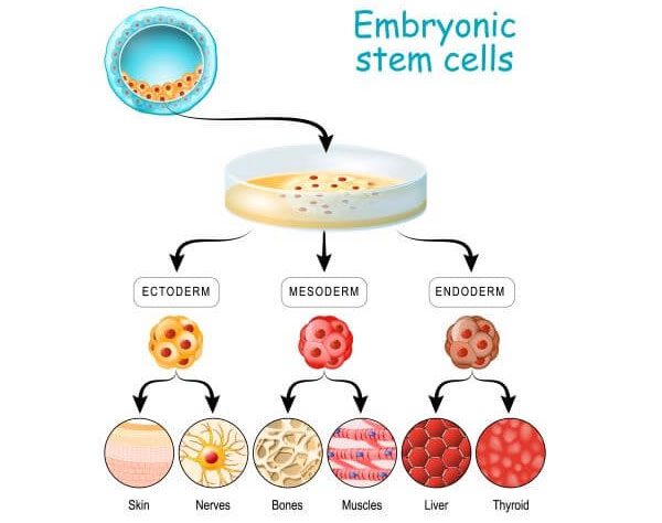
Embryonic stem cells can transform into any cell type in the body.
About a week after cultivation, Dr. Liu observed under a microscope that the crab-eating macaque stem cells had differentiated into three cell lines that form the foundation for tissue and organ development.
By days 8 and 9, these cells began organizing into structures resembling yolk sacs, which appear in the early stages of pregnancy and nourish the embryo. “This is something that scientists have yet to achieve even with natural monkey blastocyst cultures in vitro,” the study stated.
Three monkeys that received embryos showed signs of pregnancy on ultrasound
To provide further evidence, Dr. Liu and Dr. Sun sequenced the RNA from 6,000 cells present in the monkey embryos they created. The results showed that these cells possessed genetic traits similar to those of natural blastocysts.
Some cells expressed genes related to the endoderm—the innermost lining of the respiratory and digestive tracts—while others exhibited genes associated with placental development.
By day 15 after cultivation, researchers observed structures resembling the outlines of yolk sacs and amniotic membranes. While the yolk sac provides nutrition before the placenta forms, the amniotic membrane is the outer layer surrounding the developing embryo.
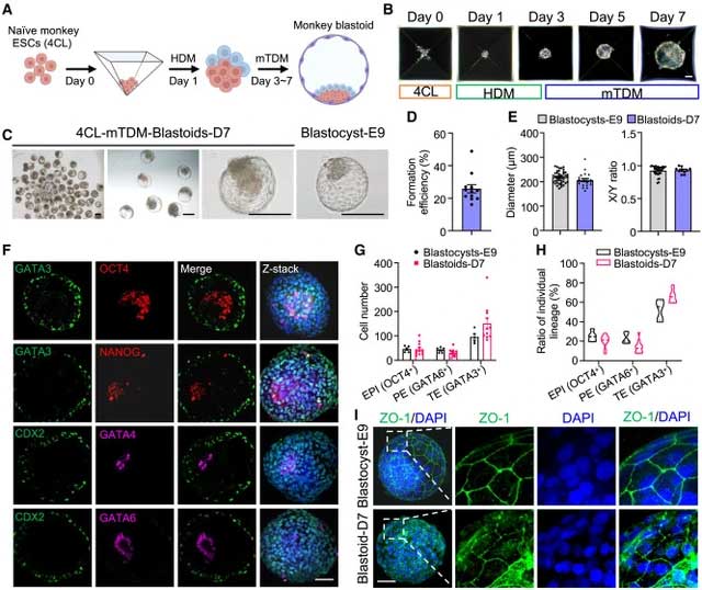
The artificial embryos developed many characteristics similar to natural monkey embryos.
The research report indicated that 5 out of 41 cultured embryos exhibited the presence of something resembling a primitive streak. This structure marks the beginning of the cell arrangement process, dividing the embryo’s layout into left-right and top-bottom orientations.
“These structures look very convincing. They seem to possess all types of cells typically found in embryos,” stated Kotaro Sasaki, an associate professor of biomedical sciences at the University of Pennsylvania School of Veterinary Medicine.
In a final step to prove they had truly created artificial monkey embryos, Dr. Liu and Dr. Sun transferred these embryos into the wombs of 8 female monkeys. After 7-10 days, 3 out of the 8 monkeys developed gestational sacs—visible fluid-filled cavities on ultrasound, marking the first sign of pregnancy.
The embryo transfer also resulted in the release of early pregnancy hormones such as progesterone and gonadotropin. However, these only persisted for a short period. Both the embryonic tissue and signs of pregnancy disappeared by day 20. Chinese scientists reported that no actual fetuses formed.
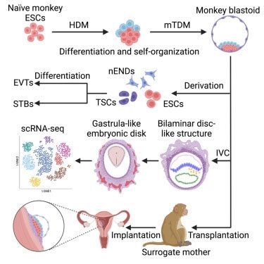
If the embryos were not transferred into the monkey uterus and were instead allowed to continue developing in a petri dish, they would continue to grow. However, at this later stage, Associate Professor Sasaki stated that the embryos had become “disorganized and no longer resembled normal embryos.”
These failures may have been due to structural or genetic abnormalities.
Why do scientists want to create monkey embryos this way?
To understand the reason, we must discuss the early days of embryonic development. In nature, after sperm meets the egg, it begins the process of embryo formation, transforming the egg into a ball that rolls down the uterus. Inside this ball, embryonic stem cells start to form.
However, once the embryo reaches its destination, it attaches to the uterine wall and disappears from view.
Scientific ethics prevent researchers from observing what happens next here, as regulations prohibit the culture of human embryos beyond 14 days in the laboratory. Other probing methods, such as ultrasound, are not sensitive enough to peer inside a human uterus during this stage.
Thus, while searching for solutions, scientists have attempted to shift to a new direction, creating embryonic structures like those developed by Dr. Liu and Dr. Sun. These can be referred to as designed artificial embryos, embryo-like structures, or pseudo-embryos—due to the fact that they will only develop for a short period without maturing into fetuses.
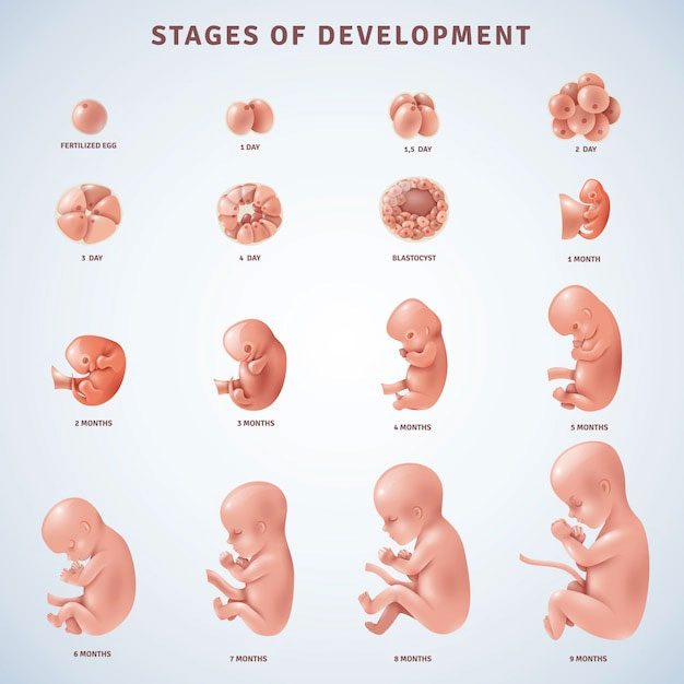
The developmental process of human embryos.
In a breakthrough in 2018, Nicolas Rivron, an embryologist at the Austrian Academy of Sciences, created mouse pseudo-embryos that developed into blastocyst-like structures by days 5 and 6 after fertilization.
In 2021, two research teams at the University of Texas and Monash University in Australia even created human pseudo-embryos that developed to day 10 after fertilization. These pseudo-embryos were created from embryonic stem cells, induced pluripotent stem cells, and skin cells.
Pseudo-embryos have allowed researchers to observe the initial processes involved in embryo formation. However, the problem with mouse pseudo-embryos is that they develop relatively differently from humans, as mice and humans are far apart on the evolutionary tree.
Meanwhile, human chimeras must be destroyed by law on the 14th day, before the appearance of neural cells. The International Society for Stem Cell Research (ISSCR) also strictly prohibits transferring these structures into humans to initiate the pregnancy process. Therefore, the emergence of mouse and human chimeras has yet to resolve the deadlock.
Now, the monkey chimeras recently created by Chinese scientists have reconciled both options. Monkeys are a species closely related to humans but are non-human primates. This is why they can implant their chimeras into mother monkeys without encountering ethical principles.
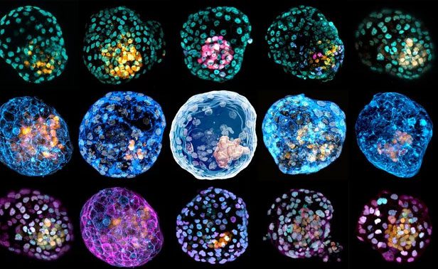
Structure of a human chimera developed by Monash University, Australia.
Scientists hope these cell clusters will provide the first clues about the development process of embryos, including the formation of diseases, congenital disabilities, and what might lead to miscarriages.
“Since monkeys have a close evolutionary relationship with humans, we hope that studying these models will help us understand human embryonic development better, including shedding light on some causes of early miscarriages,” Dr. Liu said.
What if the chimera actually develops into a fetus?
It is important to note that the report from Dr. Liu and Dr. Sun’s research indicates that their chimera died after 20 days of development. This means these cell clusters are still merely models and not actual embryos.
However, this still provides clear evidence that the field of creating chimeras from stem cells is progressing. The chimeras are becoming more similar to real embryos, and in this study, they even produced signs of pregnancy in female monkeys.
“These chimeras can initiate the pregnancy process, or at least activate the hormonal system of female monkeys so that they think they are pregnant,” Hank Greely, director of the Center for Law and the Biosciences at Stanford University School of Medicine, stated. “This is evidence that this technology has the potential to create a real monkey offspring.“
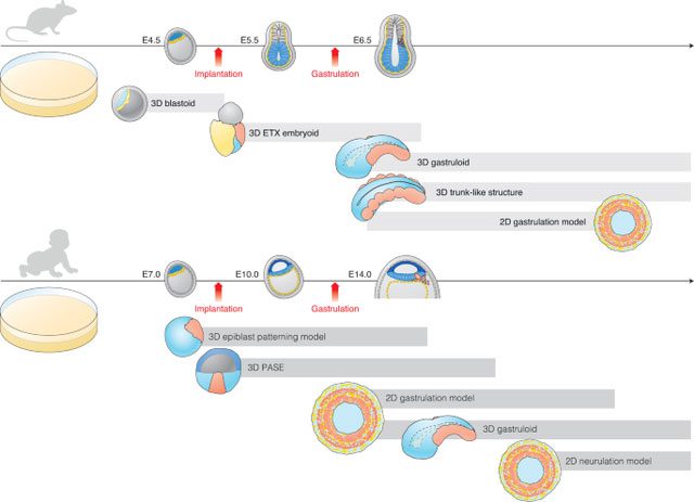
Scientists have developed mouse and human chimeras. They predict the first mouse born from a chimera will appear within the next five years.
For Rivron, he believes that sooner or later, chimeric technology will approach creating embryos that perfectly resemble natural embryos. Rivron predicts the first mouse born from a chimera will appear within the next five years.
Of course, the same cannot be done with humans. Any process that pushes human chimeras beyond the 14-day developmental milestone is considered unethical. If a human chimera were to develop into a fetus, it could lead to children with severe disabilities or genetic disorders.
Therefore, hopes are pinned on the artificial chimeras that Chinese scientists are developing.
“I think what we really want to know is: Can an embryonic model create a living organism? If it can, then it should be considered an embryo, not just a chimera. If it cannot, then it should not be treated as an embryo,” Greely commented.
To answer these questions, he advocates for scientists to conduct more experiments. However, he emphasizes that they must proceed with caution. “I think we should take things slowly to ensure we are doing it right,” Rivron remarked.
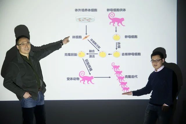
The two authors of the new study, Dr. Zhen Liu, a developmental biologist at the Institute of Neuroscience, Chinese Academy of Sciences, Shanghai (CAS), and Dr. Qiang Sun, a neuroscientist and director of the Non-Human Primate Research Facility at CAS.
For their part, Chinese scientists also acknowledge the ethical concerns surrounding their new research. They call for the international scientific community to seriously discuss this issue to find the right direction for the development of artificial embryos from human stem cells, as well as animal stem cells.
