More than 100 years ago, German biologist Richard Semon coined the term “engram,” referring to the physical or chemical changes in the brain that occur when memories are formed.
The Process of Memory Formation
Scientists now understand that when a new experience leads to the formation of long-term memory in our minds, specific neurons encode the details that, when activated, allow us to recall them.
These crucial neurons are known as engram cells. Thanks to advancements in technology over the past few years, scientists can now zoom in on the details within engram cells at higher resolutions and closely monitor the memory formation process.
According to a research article published in Nature Neuroscience, Professor Li-Huei Tsai’s team from the Massachusetts Institute of Technology (MIT) revealed for the first time that epigenetic changes occur in the genetic material of engram cells at various stages of memory formation. Large-scale changes in the 3D structure of the genome regulate the expression of specific genes related to memory storage.
To track engram cells, scientists shifted their research to specially modified gene mice. Their genome was tagged with fluorescent proteins, causing the cells to glow when they expressed the Arc gene, which is associated with memory formation.
The researchers used mild shocks to the feet to create fear memories at specific locations in mice and in the hippocampus of their brains, a crucial region for learning and long-term memory. The engram cells encoding the memory were observed to glow yellow under fluorescence. Subsequently, the researchers analyzed the glowing cells in detail hours and days after the memory was formed and when the memory was reactivated.
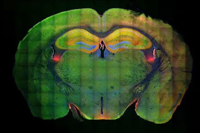
A slice of the entire mouse brain, showing glowing neurons during memory formation and recall.
During the encoding phase of memory, they noticed subtle changes in the structure of chromatin in the nucleus. Chromatin is formed by tightly winding long strands of DNA and proteins, and when specific regions undergo epigenetic changes to become looser, the DNA is exposed, allowing the genes on it to be more easily “read.”
However, they were surprised to discover that the loose regions were not coding gene segments; instead, they contained non-coding sequences known as enhancers. These sequences serve specific genes and help activate them.
In the following five days, the memory consolidation phase saw even more changes in the 3D structure of the surrounding chromatin, with many enhancers moving closer to the genes they serve.
Nevertheless, until this point, gene expression in the cells had not changed significantly as the researchers had anticipated, a result that, according to lead author Dr. Asaf Marco, left them disappointed.
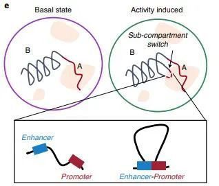
During memory formation, the 3D structure of chromatin changes, causing enhancers to move closer to the gene expression sites.
Next, the researchers returned the mice to the environment where the memory was initially formed. When the memory was resurrected, gene expression occurred rapidly. Many genes activated by enhancers were involved in the synthesis of junction proteins, leading to stronger connections between neurons forming quickly.
Dr. Marco stated: “Only then did we realize that the previous structural changes in chromatin were preparing the cells to enhance memory during the recall phase.” Another expert remarked on the memory formation process: “It’s like warming up before exercising; the engram cells are ready to activate so we can start recalling.”
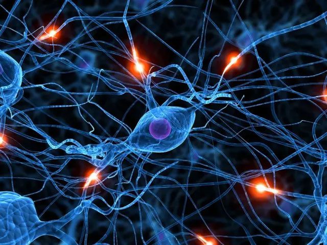
During recall, proteins related to memory storage are produced in large quantities, and connections between neurons are reinforced.
Dr. Marco concluded: “This study is the first to show that memory formation is driven by the stimulation of gene expression during the recall phase by enhancers beginning with epigenetic modifications.”
What is the Mechanism of Long-Term Memory Formation?
Scientists at Harvard Medical School have been trying to unravel this mystery for over half a century, with their findings published in the prestigious journal Nature.
Let’s turn back the clock to 1986. At that time, Professor Michael E. Greenberg, the corresponding author of this study, had just arrived at Harvard University.
In one study, he and his colleagues discovered that once a neuron is activated, it starts expressing a gene named Fos for a short period. Although the Fos gene encodes a transcription factor, scientists were uncertain about its actual function, but they hypothesized it served as a marker for neuron activation.
However, the expression patterns of Fos indicated that it was likely related to some functions of neurons that affect our ability to learn and remember.
To test this idea, in this study, the scientists placed mice in a new environment and assessed the activity of key neurons in their hippocampus. Strangely, the neurons expressing the Fos gene did not cluster together after being exposed to the new environment; instead, they were scattered throughout.
Could this also affect memory formation? Subsequent research confirmed this. After inhibiting the neurons producing Fos, the mice showed significant memory deficits, getting stuck in mazes and having difficulty escaping. This also indicated that the neurons expressing Fos were indeed involved in the memory formation process.

After exposure to the new environment, the hippocampal neurons expressing the Fos gene (in red) were not concentrated but scattered throughout.
Using optogenetic methods, the scientists activated other neurons around these neurons and found that they were influenced by two types of signals between the neurons: one type of inhibitory signaling was enhanced, and the other weakened.
If the neuron itself does not express Fos, it will not exhibit similar properties. Additionally, the relationship between activating these neural signals and other neurons in the loop is also crucial. The researchers noted that Fos could be related to the plasticity of specific neural circuits. As Fos is a transcription factor, the researchers naturally considered analyzing other genes it controls.
By employing techniques like single-cell sequencing, they identified an important gene called Scg2, which affects inhibitory signaling. If the Scg2 gene in mice is inactivated, the neurons activating Fos develop defects in signal reception.
Correspondingly, the mice also encountered issues with brain waves related to learning and memory. Specifically, Scg2 encodes a neuropeptide divided into four different forms. The researchers demonstrated that neurons use these neuropeptide chains to refine signals from other neurons.
In summary, the scientists proposed a model: when exposed to something new, a small cluster of neurons in the hippocampus simultaneously expresses Fos, activates the Scg2 gene, and produces the corresponding neuropeptide. After receiving guidance from interneurons, these neurons form a coordinated loop.
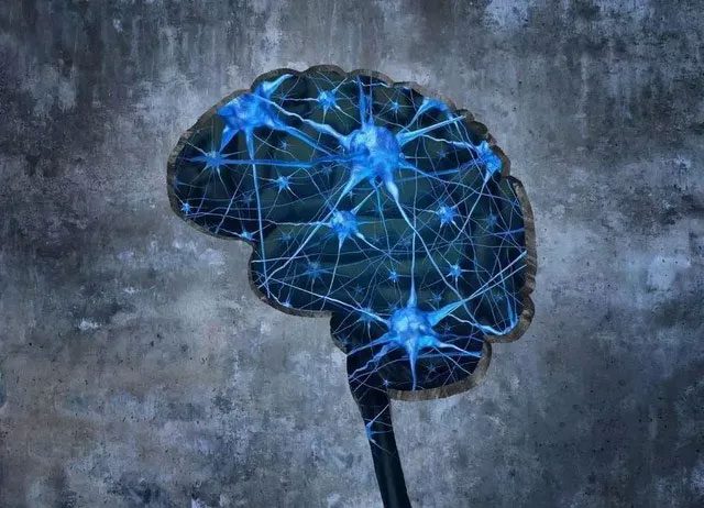
“When the neurons in the hippocampus are activated, they do not need to be connected in a specific way beforehand. The neurons between these neurons have very broad axonal branches that can connect to many cells simultaneously and transmit signals. It may be these isolated neurons that are connected in,” the research team stated.
This study provides a mechanism for long-term memory formation from a molecular perspective. Whether for basic biological research or memory-related diseases, it holds significant implications.
Why Do Some Memories Fade Away?
Information is first encoded into short-term memory, where it can be stored for a duration ranging from a few seconds to several minutes. After that, it is transferred to long-term memory through areas such as the hippocampus and eventually to various storage regions in the brain.
If two neurons continuously communicate, the efficiency of communication between them increases. This process, known as long-term potentiation, is considered a mechanism that helps memories be stored long-term, but how do some memories fade away?
Age is a factor. The hippocampus loses 5% of its nerve cells every decade, resulting in a total loss of 20% by the age of 80. This leads to a decrease in the production of neurotransmitters, such as acetylcholine, which are crucial for learning and memory. These changes seem to affect how people retrieve stored information.
Another cause of memory problems is chronic stress. Young individuals who are constantly overwhelmed by work and personal responsibilities find their bodies in a state of heightened alertness. This response develops from physiological mechanisms designed to ensure our survival during crises.
The substances produced to help reduce stress mobilize energy and increase alertness. However, with chronic stress, our bodies become flooded with these chemicals, leading to brain cell loss and an inability to form new cells, which impacts our capacity to remember new information.
Depression is another culprit. Individuals suffering from depression are at a 40% higher risk of experiencing memory problems.


