The HYBRiD technique for tissue transparency developed by the Scripps Research Institute (USA) is believed to facilitate the analysis of biological processes in the human body and diseases such as Covid-19.
Scientists at the Scripps Institute have recently announced a new tissue clearing method that can render large biological samples transparent. This method allows researchers to easily observe and study biological processes related to diseases and what occurs across various organ systems in the body more easily than ever before.
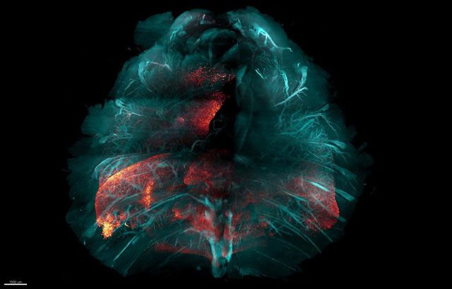
Using the HYBRiD method to observe the entire chest of a mouse infected with SARS-CoV-2 reveals that the virus’s protein appears red, while the tissue structures (lungs, blood vessels, bones) appear blue.
This method is named HYBRiD, combining elements from two previous major approaches to tissue clearing technology. HYBRiD was introduced in the scientific journal Nature Methods on March 28.
The study’s author, Dr. Li Ye, a professor of neuroscience at the Scripps Research Institute, stated: “This is a simple and widely applicable tissue clearing technique for studies on large body parts or even whole organisms.”
Tissue clearing involves using solvents to remove molecules that cause tissue to become opaque (such as fats), rendering the tissue optically transparent—while preserving most proteins and structures. Scientists often use genetically encoded or antibody-linked fluorescent markers to label active genes or other molecules in animal tissues. The tissue clearing process, in principle, allows for imaging these markers simultaneously across the entire organism.
Researchers began developing tissue clearing methods about 15 years ago, primarily aimed at tracing neural connections throughout the brain. While these methods are effective for the brain, they have proven less effective when applied to other body parts or whole organisms, which contain more challenging-to-dissolve structures.
So far, tissue clearing methods have utilized organic solvents or water-based solvents, which are better at preserving fluorescence but less effective at clearing non-brain tissues. Additionally, these methods often require labor-intensive processes and frequently involve hazardous chemicals.
Yu Wang, a research fellow in Ye’s lab who also participated in the study, mentioned: “A typical laboratory often cannot routinely and at scale use these methods.”
The new method devised by Ye and his team employs a sequential combination of organic solvents and water-based detergents, while also using water-based hydrogels to protect the molecules in the tissues that need to be preserved.
The researchers have demonstrated the ease and utility of this new method. Dr. John Teijaro, an associate professor of immunology and microbiology, has used this technique to visualize images of cells infected with SARS-CoV-2 throughout the entire chest cavity of mice.
Ye and his team are currently collaborating with their scientific partners on various applications of this new method, including tracking neural pathways in the body.


