According to information from Corney-Welsh University, scientists from the United States and Japan have successfully bred experimental mice with glowing hearts, incorporating calcium fluorescent molecules into the heart tissue of these mice, which are activated when the heart contracts.
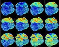 |
| Observing the glowing fetal mouse heart (Photo: VNN) |
Within the bodies of these experimental mice, there exists a gene that can self-synthesize luminescent albumin; under normal conditions, this phenomenon is typically found only in jellyfish. The luminescent DNA segment from jellyfish has been previously grafted into onions, as well as into the bodies of mice, pigs, and monkeys for research purposes.
However, Japanese scientists had to make very slight modifications to the molecules, as the fluorescence needed to be bright enough for internal organ observation. Given that the hearts of experimental mice beat up to 10 times per second, without modifications to the luminescent molecules, observation would not be feasible.
Using this method, biomedical engineers can observe the development of the heart in fetal mice without having to destroy the embryos.
In the images captured during the experiment, they were able to observe the sequential development of the two ventricles in the heart, followed by the formation of four ventricles (these represent two developmental stages on the 10th and 14th days after the embryo’s formation).
Additionally, researchers noted that during experiments on the mice, they discovered “slowing cells” that inhibit the conduction between the heart and ventricles during the early developmental stage, but these cells eventually disappear. The researchers plan to introduce this new method to other tissues, including neural tissues. They hope that in the future, this method can be applied clinically, implanting fluorescent cells into tissues that require diagnosis.
Tuyết Nhung

