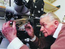
 At the Karolinska Institute (Stockholm, Sweden), alongside silent scientists, 83-year-old photographer Lennart Nilsson passionately captures images of living entities that are invisible under normal conditions.
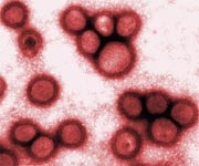
Here is the H5N1 avian influenza virus embedding its RNA molecules into a human lung cell, the world’s first three-dimensional image of H5N1. This image was obtained using a “cracking” method to break the frozen virus in liquid nitrogen, followed by observation with an ultra-powerful electron microscope.
To capture extremely dangerous viruses such as HIV, SARS, H5N1, and smallpox, Lennart Nilsson worked with inactivated viruses, which are used in vaccine research while collaborating with some of the most renowned scientists in the world. The HIV virus photo was taken in 1984 with Professor Luc Montagnier of the Pasteur Institute, while the smallpox virus was photographed in collaboration with Professor Britta Wharen of the Karolinska Institute.
The stem cells used were provided by Dr. Anders Haegerstrand of Neuronova (Sweden), extracted from the brain of volunteer patients during procedures like draining fluid from the brain or treating epilepsy.
Lennart Nilsson’s tools include a camera and the latest electron microscopes. For the past 30 years, he has closely collaborated with Japanese and German companies to order the latest types of microscopes, endoscopes, and objective lenses.
The 1965 issue of Life magazine featuring the first-ever fetal photo taken by Lennart Nilsson (in 1953) on its cover made him famous worldwide. Eight million copies of Life were sold out thanks to this photo along with the article “Life Before Birth.”
Among all his works, Lennart Nilsson is most pleased with the image of a fertilized egg dividing for the first time before implanting into the uterus. This marks the beginning of all life.
When Life Begins

1. Egg and sperm: The meeting.
2. Fertilized egg: A few hours after the egg and sperm meet, the egg begins to differentiate and moves toward the uterus aided by millions of tiny cilia swimming. This is the moment of all beginnings!
3. 16-week-old fetus: Measuring 16 cm in length, the fetus’s leg bones are dark, visible through the skin. The rest is in cartilage.

4. 25-week-old fetus: Measuring 43 cm, with formed fingernails, eyelids, and developed lungs, though not yet mature. The placenta is not yet fully functional.
5. 20-week-old fetus: Measuring 30 cm, half the length of a newborn. The skin is thin and wrinkled. Fingernails, eyebrows, and eyelashes have begun to grow. Eyelids are still closed.
6. Estrogen: Female sex hormone in crystalline form.
7. Testosterone: Male sex hormone in crystalline form.

1. H5N1: The green avian influenza virus is destroying a lung cell (pink). Its next step is to embed its RNA molecules into the cell.
2. Smallpox virus: The blue virus clings to the surface of a paralyzed cell. It has been eradicated worldwide through vaccination, one of the six infectious diseases that have claimed the most lives.
3. Cancer: A yellow urine cell approaches a cancer cell. The kiss of death.
4. SARS: Thousands of green viruses emerge from a pink lung cell after replication. Emerging in China in 2002, SARS was the first serious infectious disease of the 21st century, rapidly spreading worldwide in 2003, infecting over 8,000 people and causing 800 deaths.
5. HIV: Pink T-lymphocytes being replicated and attacked by the HIV virus (yellow). By 2005, the number of people living with HIV globally surpassed 40 million. Since its emergence in 1980, HIV has killed 25 million people.
6. Stem cells: Opening pathways for future healing, stem cells are isolated from brain cells extracted from volunteers.
DINH CONG THANH


