According to a study published in the scientific journal “Nature Medicine,” a team of Belgian and British scientists has successfully created miniature organs in the body using amniotic fluid. These “mini lungs” can be used to better predict the lung condition of a fetus in the womb.
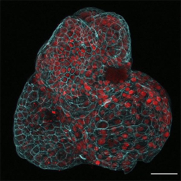
Microscopic image showing a lung organ created using cells collected from amniotic fluid. (Photo: AP).
According to reporters in Brussels, researchers from KU Leuven University in Belgium, University College London, and Great Ormond Street Hospital have used cells from amniotic fluid to culture three-dimensional cells, from which they created small organs that mimic the structure and function of the organs from which the cells were derived. Specifically, scientists have developed mini organs using cells from the kidneys, intestines, and particularly the fetal lungs. The success of the team also demonstrates that using amniotic fluid cells can help predict organ function after birth.
In the next steps, researchers will culture organs from cells taken from the amniotic fluid of fetuses with abnormal small lungs due to diaphragm defects. This type of congenital defect can lead to infant mortality, with a death rate exceeding 30%. Additionally, these mini organs can also be used for drug testing before birth, increasing efficiency and shortening treatment times for newborns.
Millions of fetal cells are found in the amniotic fluid of pregnant women, with a large portion coming from the lungs and kidneys. When ultrasound results suggest a suspicion of congenital defects, doctors often recommend amniocentesis, extracting a quantity of amniotic fluid from the mother’s body to study and detect abnormalities early.


