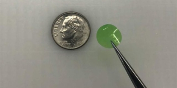
The device is made from SnS nano sheets – a type of ultra-thin material, with a thickness of only 10 nanometers – which is half the thickness of current X-ray devices and approximately 10,000 times thinner than a sheet of paper.

SnS nano sheets allow for efficient X-ray absorption. (Illustrative image).
According to scientists, this is the first time this material has been used to create a X-ray detector. The SnS nano sheets enable efficient absorption of X-rays, allowing biologists to observe cellular interactions and processes with “higher temporal resolution.” Previously, scientists could only study the results of cellular interactions but could not observe the processes in detail.
Professor Jacek Jasieniak from the Department of Materials Science and Engineering at Monash University, the lead author of the study, stated that this device provides images almost instantly. Unlike the “hard” X-rays commonly used in medicine, particularly for imaging fractures, the “soft” X-rays are used to study the structures of biological samples at the nano level, such as tissue samples.
Researchers indicated that more time is needed to explore the full potential of the device. The next step for the team will be to test different thicknesses of the device in biological cell environments. This device lays the groundwork for the development of more advanced technologies.
X-rays have contributed to some of the greatest scientific discoveries made in the 20th century, including verifying the wave nature of photons, quantifying the structure of materials, and capturing images of tissues and structures within living organisms.
The study was published in the journal Advanced Functional Materials.


