Hydrocele is a condition characterized by the accumulation of fluid, blood, or pus between the two layers of the tunica vaginalis, the membrane surrounding the testicle. Hydrocele is a symptom that can arise from various causes.
 |
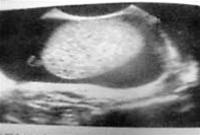
| Ultrasound image of hydrocele (Photo: TTO) |
The tunica vaginalis is a layer formed by the peritoneum that descends during the descent of the testicles. Initially, the peritoneum descends into the scrotum as a tube called the processus vaginalis, which eventually gets sealed off at birth.
The tunica vaginalis consists of two layers: the visceral layer that adheres closely to the testicle, and the parietal layer forming the outer wall of the visceral layer. Between these two layers is a thin layer of fluid that allows the testicle to move freely up and down.
There are numerous causes of fluid accumulation in the tunica vaginalis, including infections caused by bacteria affecting the reproductive and urinary systems such as E. coli, gonorrhea, syphilis, various strains of streptococcus, staphylococcus, and particularly tuberculosis bacteria. It can also result from parasitic infections such as filariasis or fungal infections; systemic diseases that lead to hydrocele include heart failure, cirrhosis with ascites, nephrotic syndrome, and more.
Other causes include cancer, trauma, or simply as a consequence of inflammatory reactions in the testicle and epididymis.
Signs and Symptoms of Hydrocele
Pain: Patients may experience severe pain, cramping, or a dull, continuous ache in the scrotum and groin area.
Swelling of the scrotum: The scrotum may appear enlarged and drooping, with tight, shiny skin, but the testicles do not descend, and the skin pinch test is negative (-). A flashlight shone onto the scrotum can easily penetrate the light.
The testicle may swell, be painful, or feel as hard as a rock.
In cases of epididymitis or orchitis, the tunica vaginalis can also become irritated and secrete fluid, leading to severe pain and swelling of the testicle and epididymis.
In cases of minimal fluid accumulation, it may be challenging to detect through clinical signs. Therefore, ultrasound is essential for quick and accurate detection. On ultrasound, a fluid layer thicker than 5mm is significant (if less, it may be normal physiological fluid).
Hematic fluid: Trauma or cancer.
Purulent fluid: Acute inflammation caused by bacteria.
Lemon-yellow fluid: Typically exudate due to systemic diseases, which may be due to tuberculosis or cancer.
Chylous fluid: Caused by filariasis, when chylous fluid is present, patients exhibit very characteristic clinical signs of filariasis (elephantiasis, chyluria).
Treatment of Hydrocele
For bacterial infections of the urinary, reproductive, and sexual systems, medications such as cefuroxime 2g/day, cefixime 400-800mg/day, ceftriaxone 1g/day, or quinolone groups like ofloxacin, sparfloxacin, or rotifloxacin may be prescribed by a physician.
In cases of tuberculosis, which is usually secondary, treatment should follow the prescribed regimen.
For malignant causes, surgical intervention, radiation therapy, or chemotherapy may be considered based on the specific case.
In cases of parasitic infections, such as filariasis, diethylcarbamazine is used.
Aspiration of the tunica vaginalis fluid serves both as a diagnostic procedure and a treatment technique for cases with significant fluid accumulation.
Hydrocele presents as a symptom of various causes and clinical scenarios. Treatment must be based on the underlying cause for optimal effectiveness. The treatment goal is not only to alleviate symptoms in the tunica vaginalis but also to preserve testicular function and maintain sexual and reproductive health for the patient afterward.


