To create a human heart, researchers must replicate the unique structures that make up the heart. This design involves establishing spiral connections of cardiac tissue, which create a twisting motion as the heart beats.
For a long time, scientists have hypothesized that the twisting motion is crucial for pumping blood in large volumes, but proving this has been challenging, partly because creating a heart with different shapes and rational arrangements is a significant challenge.
Biomedical engineers from the Harvard John A. Paulson School of Engineering and Applied Sciences (SEAS) have developed the first biologically integrated model of the human ventricle with spirally connected beating heart cells, demonstrating that the actual connection of cardiac tissue significantly increases the volume of blood the ventricle can pump with each contraction.
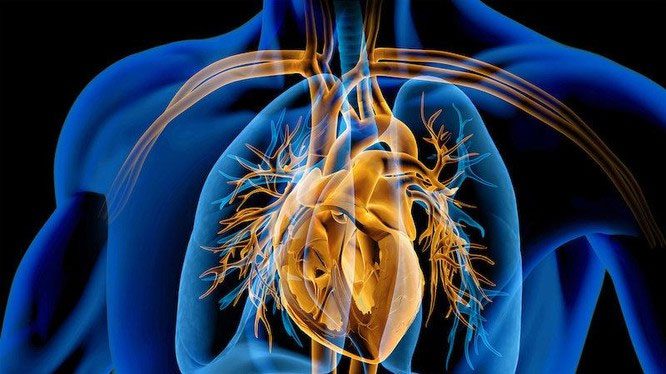
Illustration of a cross-section of the human heart. (Photo © matis75 / stock.adobe.com)
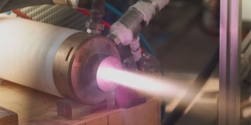
This advancement was achieved using a new additive manufacturing method. The Focused Rotational Jet Spinning (FRJS) technique allows for the high-throughput production of spiral fibers with diameters ranging from a few micrometers to hundreds of nanometers.
Developed at SEAS by the Kit Parker Disease Biophysics Group, FRJS directs the alignment of cells, enabling the formation of controlled tissue design structures.
The study was published in the journal Science.
Parker, a professor of Biomedical Engineering and Applied Physics at SEAS, stated: “This work represents a significant step forward in the process of organ fabrication and brings us closer to the ultimate goal of building a human heart for transplantation.”
This work originates from a centuries-old mystery. In 1669, English physician Richard Lower, who considered John Locke one of his colleagues and King Charles II among his patients, first noted the spiral arrangement of cardiac muscles in his work Tractatus de Corde.
In the following three centuries, doctors and scientists built a more comprehensive understanding of the heart’s structure, but research into the purpose of these spiral muscles remained challenging.
In 1969, Edward Sallin, former chair of the Biology Department at the University of Alabama at Birmingham, argued that the heart’s spiral arrangement is crucial for achieving a high ejection fraction—the percentage of blood the ventricle pumps with each contraction.
John Zimmerman, a postdoctoral researcher at SEAS and the first co-author of the paper, indicated that the goal was to create a model to test Sallin’s hypothesis and investigate the relative importance of the heart’s spiral structure.
To test Sallin’s theory, SEAS researchers used the FRJS system to control the alignment of the spiral fibers, on which heart cells could grow.
The first step of FRJS operates like a cotton candy machine—a liquid polymer solution is loaded into a reservoir and pushed out through a small hole by centrifugal force as the device spins. As the solution exits the reservoir, the solvent evaporates, and the polymers solidify into fibers.
Then, a focused airflow controls the direction of the polymer fibers as they wind around a collector. The research team discovered that by angling and rotating the collector, the fibers in the stream would align and twist around the collector as it spun, mimicking the twisted structure of cardiac muscle.
The arrangement of the fibers can be adjusted by changing the angle of the collector.
Huibin Chang, a postdoctoral researcher at SEAS and a co-author of the paper, remarked: “The human heart truly has many layers of muscle arranged in spirals with different angles. With FRJS, it is possible to recreate these complex structures very accurately, forming a single ventricular structure and even four chambers.”
Unlike 3D printing, which slows down as materials become smaller, FRJS can quickly spin fibers at a micro-scale—or about fifty times smaller than a human hair.
Artificial ventricle, a significant advancement in the process of creating biological organs. (Video by Harvard John A. Paulson School of Engineering and Applied Sciences).
This is crucial when building a heart from scratch. Collagen, an extracellular matrix protein in the heart, also has a diameter of one micrometer. It would take over 100 years to 3D print each collagen part of the human heart at this resolution. FRJS can accomplish this in one day.
After spinning, the ventricles are seeded with cardiac muscle cells from mice or human stem cell-derived cardiac muscle cells. Within about a week, several layers of beating tissue cover the scaffold, with cells developing according to the alignment of the underlying fibers. The beating ventricles mimic the twisting or wringing motion seen in the human heart.
Researchers compared the deformation of the ventricles, the speed of electrical signal transmission, and the ejection fraction between ventricles developed from spiral fibers and those developed from circumferential fibers. Tests showed that the spiral-aligned tissue performed better than the circumferential-aligned tissue.
The research team also demonstrated that this process could be scaled up to the size of a real human heart and even larger, reaching the size of a whale’s heart.
