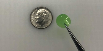
The transplantation of small human brain tissues into mouse brains has reached a new milestone. In the future, this could be applied to treat severe brain injuries.
According to Science Alert, this new research was published in the journal Cell Stem Cell. The results show that the small human brain tissues not only integrate with the surrounding mouse brain tissue but also that the neurons in the organ begin to react to visual stimuli. Remarkably, this integration occurs within just three months of transplantation.
Dr. H. Isaac Chen, a neuroanatomist at the University of Pennsylvania, stated: “We did not expect the level of functional integration to occur so early. Many previous studies involving the transplantation of human neurons into rodent species took 9-10 months and still did not achieve completion.”

Previous studies on the transplantation of human neurons into rodents did not show results as quickly as this study. (Image: Newatlas).
Research Process
Organoids are 3D tissues derived from human embryonic stem cells or pluripotent stem cells. The process of transplanting human cortical organoids into the brains of rodents is becoming increasingly sophisticated.
Previously, individual neurons were used. However, recently, researchers have successfully transplanted human cortical organoids into the brains of both juvenile and adult mice. These organoids integrate with the surrounding tissue and show signs of activity.
Currently, Dr. Chen and his team are taking the next step by transplanting human brain tissue into adult mice with significant cortical damage to see if there is also functional integration.
Dr. Chen said: “We are not only focusing on the transplantation of individual cells but also on the transplantation of tissue. Brain organoids have a structure similar to that of the brain. We can examine individual neurons within this structure to gain a deeper understanding of the integration of transplanted organoids.”
To develop the human “mini-brain,” researchers used genetically modified induced pluripotent stem cells to express green fluorescent protein (GFP), a protein composed of 238 amino acids.
Induced pluripotent stem cells are created from adult stem cells through a process of reprogramming the genome. They can develop into various types of cells. The green fluorescent protein enables the brain organoids to exhibit fluorescence.
These stem cells were developed into human neurons over approximately 80 days. Subsequently, researchers began to transplant them into the brains of 10 adult male mice.
First, they created a cavity the size of an organ (about 2 mm in diameter) in the brain of each mouse. This cavity represents severe brain injury. Then, they inserted the organoid and sutured the wound to allow it to heal.
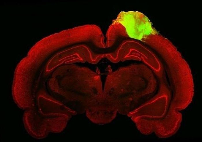
Histological image of a mouse brain with implanted human cortical organoid (green). (Image: Cell Stem Cell).
Response to Light
To observe how the organoids integrate with the brain after healing, researchers injected fluorescently tagged viruses into the eyes of the mice. They then monitored the neuronal connections from the retinas of the mice.
Subsequently, when flashing light and alternating black and white bar images were projected into the mice’s eyes, researchers used electrodes to investigate the activity within the organoid. Approximately 25% of the human neurons responded to light stimuli.
“We observed a significant number of neurons in the organoid responding to specific light directions. This indicates that the neurons in this organoid can not only integrate with the visual system but also apply specific functions of vision,” Dr. Chen noted.
The experiment was limited to three months due to immune suppression to prevent the mice’s bodies from rejecting the human tissue. By the end of the experiment, the mice had died gently.
Due to the short duration, the human neurons may not have fully matured. This explains why the responsiveness of the neurons was not high.
However, the results can still be utilized to improve future trials. The research team recommends using genetically immunosuppressed rodents for long-term studies.
“Neural tissues have the potential to regenerate damaged brain areas. Now, we want to understand how organoids can be used in other regions of the cortex, not just the visual cortex. We want to explore what allows neuron organoids to integrate with the brain, which could enable us to better control this process and make it happen faster,” Dr. Chen concluded.


